- Details
Photo By Rebekah L. Holt, eQuest Photography
What About Horse Toe Evolution?
By Rebekah L. Holt
 According to Darwin’s ideas on equine origins, the ancestor of the horse, millions of years ago, was a five-toed, fox-sized creature.1 Through random change, blind selection, and almost endless time, the original five toes fell by ones and twos, as the ancestors of the horse grew from rodent to equine.
Evolutionists teach that the hoof remains the sole toe of perfected horse evolution. The theory also proclaims that other existing parts of the leg, specifically the chestnut, the splint bones and the ergot, are all vestigial (leftover, useless) remains of horse evolution’s missing toes.
So what is the truth about horse toes? According to Genesis chapter 1, the equine kind2 was masterfully created on the sixth day. God in His skilful design made the equine and its foot like no other.
By studying each member of the ‘toe vestiges,’ we can discover fingerprints of the Divine Creator and gather evidence to refute Darwin’s false assumptions.
The Hoof
The hoof, or equine foot, is vital to a horse’s existence. This horny structure is located on the very end of a horse’s leg. Hooves play such a crucial role in the health and well-being of the horse that the old adage, ‘No hoof, no horse’ is true.
To study the equine foot uncovers a structural wonder which causes farrier, veterinarian, and layman alike to marvel. Here are just some of the tasks the hoof is designed to perform:
 Imagine the horse’s hooves as suction cups that compress with each step to absorb shock while gripping the surface to aid traction. With each step, the inner structures of the hoof work like a pump, compress-and-release, shooting life-giving blood back up the leg and to the heart. Hundreds of microscopic, straw-like tubules serve to reinforce the hoof wall’s strength and draw ground moisture with capillary action to hydrate the hoof wall.
If even one part of the horse’s foot is out of place, the result is lameness, often chronic or painfully disabling.
Splint Bones
Hailed as ‘vestigial’ or useless leftovers of evolution, splint bones actually are far from useless bits of bone. Splints are two icicle-shaped bones found on the back of each leg. They support the carpal joints (front knee) on either side of the cannon bone, and the tarsal (or hock) joints on the rear leg. In addition to assisting weight support, splints form a vital ‘groove’ and protection for ligaments and tendons that enable equine locomotion.10
 Dr. Doug Butler, a leading authority on farrier science, wrote concerning splint bones:
‘The function of the splint bones is to protect the tendons and ligaments and especially the blood vessels and nerves which pass down the back of the leg. They also provide a greater bearing surface for weight by supporting a portion of the carpal bones of the knee joint. They are necessary, not vestigial.’ [Emphasis added]
Cases have been reported of multi-toed (polydactyl) horses with extended splint bones as extra toes.11 There are skeletons of horses with splint bones extending as two extra toes. Does this prove Darwin’s theory of a transitional creature evolving into a horse? Certainly not! It only illustrates that a totally equine skeleton had extra long splint bones supporting its fossilized leg that at one time supported a member of the equine kind. The modern equine’s regressing splints suggest only that the genetic material for extended splints has been lost. Regression, shortening or loss of toes, is not helpful to the progressive evolutionary theory, which requires an overall increase in genetic complexity.
The Chestnut
 Everyone wonders what the chestnut really is. Science has not yet satisfactorily explained this little mystery. It is popularly believed that the chestnut has shrunk from a toe in the horse’s ancestor into a horny little growth inside of the horse’s leg. While that may be so (devolution is quite possible in a creation model, and is unhelpful to evolution, as shown above), it seems premature to call it vestigial. A respected veterinarian handbook states:
‘Contrary to popular belief, they [chestnuts] do not represent the vestiges of missing digits.’12
Chestnuts are called the ‘fingerprints’ of horses; each is individual in shape and texture. They can serve as identification just like our fingerprints do.13
It’s largely believed that chestnuts are scent glands (llamas have such glands in a similar place, thought to make alarm pheromones). They carry the strong trademark aroma of ‘horse.’14
Photo note: Chestnuts and ergots are unique to each horse and can be used for identification. The photos show chestnut and ergot variations of the forelimbs of three American Quarter Horse mares. The mare to the far right did not have an ergot growth—these, and chestnuts, are commonly absent in all breeds.
The Ergot
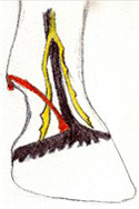
 The ergot, a small, bony growth located on the back of the fetlock joint, is considered the last of Darwin’s missing horse toes. It too is taught as a vestigial leftover of the evolving horse series. While the ergot may seem to be a useless little bump, it actually is an anchoring point for the ergot ligament—the most superficial (close to the surface) of the ligaments found on a horse’s leg.15
Illustration Notes: Orange depicts ergot ligament and ergot. Yellow depicts nerves. Black depicts the artery.
Conclusion
A recipe must have a chef, a portrait must have a painter, and a creature must have a Creator. As Christians, we can rejoice that humans and all other creatures are not remnants of thoughtless evolution, but creatures skillfully designed by our Lord and Creator.
As Revelation 4:11 declares, ‘Thou art worthy, O Lord, to receive glory and honour and power: for thou hast created all things, and for thy pleasure they are and were created.’
References
- The fossil of this supposed ancestor has been called Eohippus (dawn horse) but its original (and proper) name is Hyracotherium, reflecting its similarity not to horses, but to the living hyrax, or rock badger, aka coney. It may well be an ancestor of the hyrax.
- This most likely was the ancestor of today’s horses, zebras and asses, which can all interbreed.
- Giffin, James M., M.D. & Tom Gore, D.V.M., Horse Owner’s Veterinary Handbook, Second Edition, Howell Book House, New York, 1998, page 307.
- Butler, Doug. PhD., The Principles of Horseshoeing II, Doug Butler Publication, Maryville, Missouri, 1985, page 121.
- Ref. 4, page 120.
- Bertram JE, Gosline JM, Fracture Toughness Design in Horse Hoof Keratin, The Journal of Experimental Biology 125(1):29–47, September 1986.
- Pollitt, Christopher C., BVSc, Ph.D., The Anatomy of the Inner Hoof Wall, The International Equine Research Center, The Farrier and Hoof Care Research Center, as at 14 June 2008.
- Ref. 3, page 261.
- Ref. 4, pages 138–139.
- Ref. 2, page 100.
- Carstanjen B, Abitbol M, and Desbois C, Bilateral Polydactyly in a Foal, Journal of Veterinary Science 8(2):201–203, June 2007.
- Ref. 3, page 87.
- Hadden, Will A., III, D.V.M., Horseman’s Veterinary Encyclopedia, The Lyons Press, Guilford, Connecticut, 2005, page 169.
- Wolfrom, Glen, Horse Chestnuts, Creation Matters 3(4):5, July–August 1998.
- Ref. 4, page 116.
Originally Published by Creation Ministries International
Photos by Rebekah L. Holt, eQuest Photography
Illustrations by Rebekah L. Holt


